The Soeller Lab at the University of Bern
The Soeller Lab focuses on cardiac physiology while also having a strong interest in optical super-resolution imaging.
New MINFLUX publications
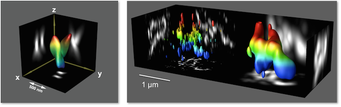
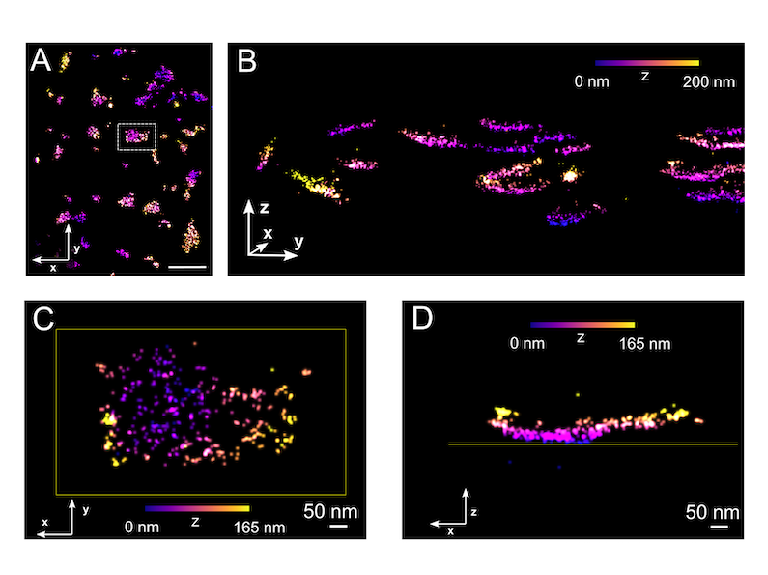
We have recently published two studies using MINFLUX microscopy to image receptors in cardiac myocytes at full molecular resolution.
For details see our study in Nature Communications MINFLUX microscopy resolves subunits of the cardiac ryanodine receptor... and the companion methods paper in ACS Photonics Optimizing Effective Labeling Efficiency in MINFLUX 3D DNA-PAINT Microscopy .... See also our news items here and here.
Investigation of nanoscale structure-function relationships
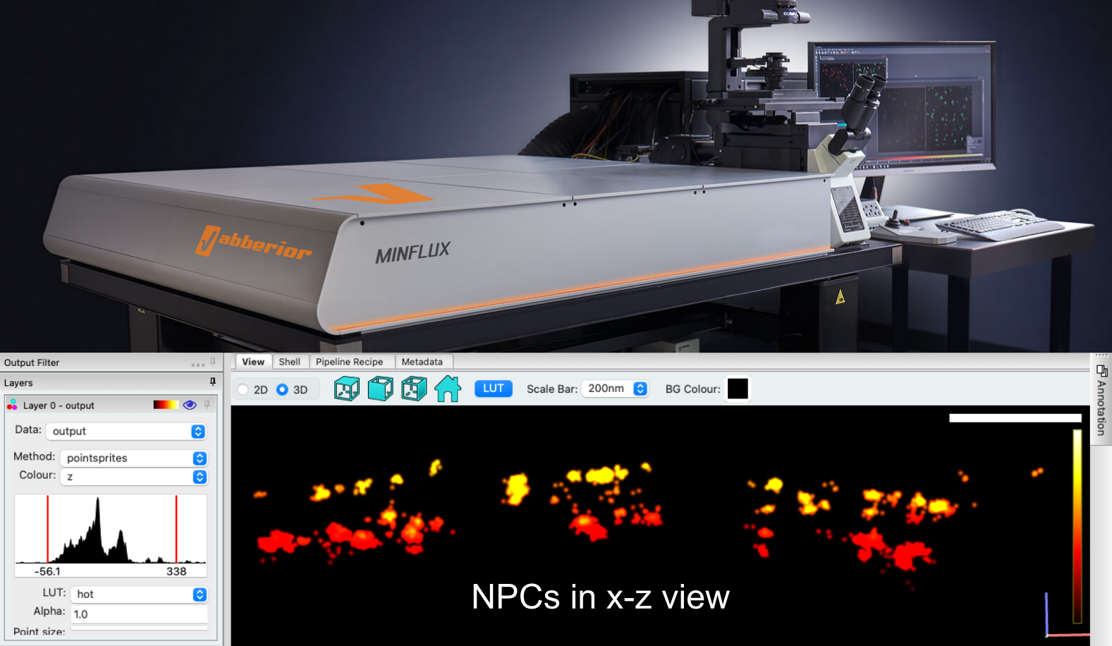
Physiology Bern is now hosting a fully speced MINFLUX super-resolution system. We are very excited to introduce truly molecular resolution optical imaging to Bern. The system has been in place since late 2023 and we have been working up the technology for wider use by both us and other users. This latest super-resolution technology allows us to carry out molecular structure-function relationships at unprecedented spatio-temporal resolution.
Openings
We are always happy to talk to interested PhD students or thoese interested in postdoctoral work. For details consult our openings page. Please contact us for any inquiries.
The Laboratory
Our laboratory is based at the Institute of Physiology at the University of Bern.
We use advanced imaging approaches to address a variety of questions in cardiac physiology. Imaging in general, and fluorescence imaging in particular, is playing an increasingly important role in Biophysics and Biology. Our ability to come up with mechanistic descriptions of physiological processes depends to a large extent on our ability to see the components of a cell, an organism, etc. Our laboratory therefore applies and develops state-of-the-art microscopy methods to improve our understanding of the world around us.

Physiology
Our work is motivated by the goal to improve our knowledge of the physiology and biophysics of cardiac calcium regulation. Our primary focus is on cardiac ventricular muscle with a unifying theme to elucidate the relationship between nanoscale cell morphology and calcium signalling.
Advanced Imaging
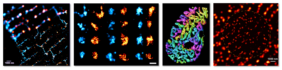
Our understanding of how biological systems work is dependent on the ability to see these systems, ideally with a resolution that approaches subcellular and even molecular scales. This has become possible by rapid advances in fluorescence imaging. The holy grail of advanced imaging is fully quantitative microscopy, that allows us to count molecules in situ, fully spatially resolved, so that we can distinguish different populations, provide molecular statistics, and similar quantitative measures that link form and function. Such quantitative molecular imaging is now becoming a practical reality with the latest imaging modalities.